Example of Microscopy and Microanalysis format
Recent searches

Example of Microscopy and Microanalysis format Example of Microscopy and Microanalysis format Example of Microscopy and Microanalysis format Example of Microscopy and Microanalysis format Example of Microscopy and Microanalysis format Example of Microscopy and Microanalysis format Example of Microscopy and Microanalysis format Example of Microscopy and Microanalysis format Example of Microscopy and Microanalysis format Example of Microscopy and Microanalysis format Example of Microscopy and Microanalysis format Example of Microscopy and Microanalysis format Example of Microscopy and Microanalysis format Example of Microscopy and Microanalysis format Example of Microscopy and Microanalysis format Example of Microscopy and Microanalysis format Example of Microscopy and Microanalysis format Example of Microscopy and Microanalysis format Example of Microscopy and Microanalysis format Example of Microscopy and Microanalysis format Example of Microscopy and Microanalysis format Example of Microscopy and Microanalysis format Example of Microscopy and Microanalysis format Example of Microscopy and Microanalysis format Example of Microscopy and Microanalysis format Example of Microscopy and Microanalysis format Example of Microscopy and Microanalysis format Example of Microscopy and Microanalysis format Example of Microscopy and Microanalysis format Example of Microscopy and Microanalysis format
Sample paper formatted on SciSpace - SciSpace
This content is only for preview purposes. The original open access content can be found here.
Look Inside
Example of Microscopy and Microanalysis format Example of Microscopy and Microanalysis format Example of Microscopy and Microanalysis format Example of Microscopy and Microanalysis format Example of Microscopy and Microanalysis format Example of Microscopy and Microanalysis format Example of Microscopy and Microanalysis format Example of Microscopy and Microanalysis format Example of Microscopy and Microanalysis format Example of Microscopy and Microanalysis format Example of Microscopy and Microanalysis format Example of Microscopy and Microanalysis format Example of Microscopy and Microanalysis format Example of Microscopy and Microanalysis format Example of Microscopy and Microanalysis format Example of Microscopy and Microanalysis format Example of Microscopy and Microanalysis format Example of Microscopy and Microanalysis format Example of Microscopy and Microanalysis format Example of Microscopy and Microanalysis format Example of Microscopy and Microanalysis format Example of Microscopy and Microanalysis format Example of Microscopy and Microanalysis format Example of Microscopy and Microanalysis format Example of Microscopy and Microanalysis format Example of Microscopy and Microanalysis format Example of Microscopy and Microanalysis format Example of Microscopy and Microanalysis format Example of Microscopy and Microanalysis format Example of Microscopy and Microanalysis format
Sample paper formatted on SciSpace - SciSpace
This content is only for preview purposes. The original open access content can be found here.
open access Open Access

Microscopy and Microanalysis — Template for authors

Categories Rank Trend in last 3 yrs
Instrumentation #57 of 128 up up by 21 ranks
journal-quality-icon Journal quality:
Good
calendar-icon Last 4 years overview: 454 Published Papers | 1268 Citations
indexed-in-icon Indexed in: Scopus
last-updated-icon Last updated: 26/06/2020
Related journals
Insights
General info
Top papers
Popular templates
Get started guide
Why choose from SciSpace
FAQ

Related Journals

open access Open Access
recommended Recommended

Taylor and Francis

Quality:  
High
CiteRatio: 9.0
SJR: 0.984
SNIP: 2.03
open access Open Access

Hindawi

Quality:  
High
CiteRatio: 4.1
SJR: 0.399
SNIP: 1.108
open access Open Access
recommended Recommended

IEEE

Quality:  
High
CiteRatio: 6.0
SJR: 0.681
SNIP: 1.481
open access Open Access
recommended Recommended

IEEE

Quality:  
High
CiteRatio: 6.1
SJR: 0.82
SNIP: 1.928

Journal Performance & Insights

Impact Factor

CiteRatio

Determines the importance of a journal by taking a measure of frequency with which the average article in a journal has been cited in a particular year.

A measure of average citations received per peer-reviewed paper published in the journal.

3.414

28% from 2018

Impact factor for Microscopy and Microanalysis from 2016 - 2019
Year Value
2019 3.414
2018 2.673
2017 2.124
2016 1.891
graph view Graph view
table view Table view

2.8

3% from 2019

CiteRatio for Microscopy and Microanalysis from 2016 - 2020
Year Value
2020 2.8
2019 2.9
2018 2.4
2017 1.2
2016 1.2
graph view Graph view
table view Table view

insights Insights

  • Impact factor of this journal has increased by 28% in last year.
  • This journal’s impact factor is in the top 10 percentile category.

insights Insights

  • CiteRatio of this journal has decreased by 3% in last years.
  • This journal’s CiteRatio is in the top 10 percentile category.

SCImago Journal Rank (SJR)

Source Normalized Impact per Paper (SNIP)

Measures weighted citations received by the journal. Citation weighting depends on the categories and prestige of the citing journal.

Measures actual citations received relative to citations expected for the journal's category.

0.521

15% from 2019

SJR for Microscopy and Microanalysis from 2016 - 2020
Year Value
2020 0.521
2019 0.611
2018 0.437
2017 0.292
2016 0.31
graph view Graph view
table view Table view

0.799

19% from 2019

SNIP for Microscopy and Microanalysis from 2016 - 2020
Year Value
2020 0.799
2019 0.988
2018 0.694
2017 0.274
2016 0.279
graph view Graph view
table view Table view

insights Insights

  • SJR of this journal has decreased by 15% in last years.
  • This journal’s SJR is in the top 10 percentile category.

insights Insights

  • SNIP of this journal has decreased by 19% in last years.
  • This journal’s SNIP is in the top 10 percentile category.

Microscopy and Microanalysis

Guideline source: View

All company, product and service names used in this website are for identification purposes only. All product names, trademarks and registered trademarks are property of their respective owners.

Use of these names, trademarks and brands does not imply endorsement or affiliation. Disclaimer Notice

Cambridge University Press

Microscopy and Microanalysis

Microscopy and Microanalysis publishes original research papers in the fields of microscopy, imaging, and compositional analysis. This distinguished international forum is intended for microscopists in both biology and materials science. The journal provides significant articl...... Read More
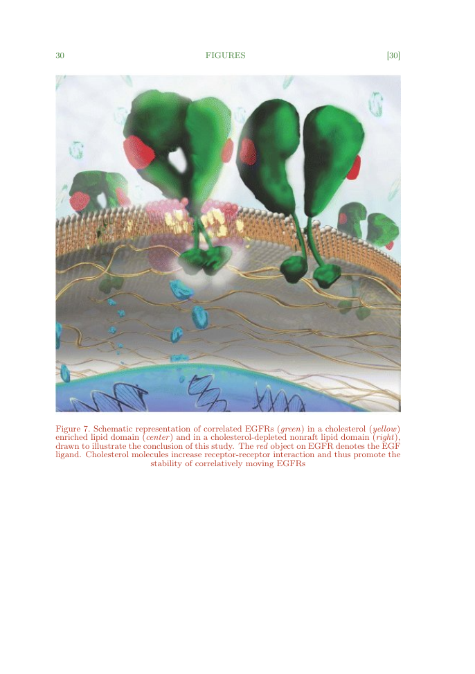
Instrumentation

Physics and Astronomy

i
Last updated on
25 Jun 2020
i
ISSN
1431-9276
i
Impact Factor
High - 2.124
i
Acceptance Rate
Not provided
i
Frequency
6 issues per year
i
Open Access
Yes
i
Sherpa RoMEO Archiving Policy
Green faq
i
Plagiarism Check
Available via Turnitin
i
Endnote Style
Download Available
i
Bibliography Name
unsrt
i
Citation Type
Author Year
(Blonder et al., 1982)
i
Bibliography Example
Blonder, G. E., Tinkham, M., and Klapwijk, T. M. 1982. Transition from metallic to tunneling regimes in super-conducting microconstrictions: Excess current, charge imbalance, and supercurrent conversion. Phys Rev B., 25(7):4515–4532.

Top papers written in this journal

Journal Article DOI: 10.1017/S1431927611000055
A review of strain analysis using electron backscatter diffraction.
Stuart I. Wright1, Matthew M. Nowell1, David P. Field2

Abstract:

Since the automation of the electron backscatter diffraction (EBSD) technique, EBSD systems have become commonplace in microscopy facilities within materials science and geology research laboratories around the world. The acceptance of the technique is primarily due to the capability of EBSD to aid the research scientist in u... Since the automation of the electron backscatter diffraction (EBSD) technique, EBSD systems have become commonplace in microscopy facilities within materials science and geology research laboratories around the world. The acceptance of the technique is primarily due to the capability of EBSD to aid the research scientist in understanding the crystallographic aspects of microstructure. There has been considerable interest in using EBSD to quantify strain at the submicron scale. To apply EBSD to the characterization of strain, it is important to understand what is practically possible and the underlying assumptions and limitations. This work reviews the current state of technology in terms of strain analysis using EBSD. First, the effects of both elastic and plastic strain on individual EBSD patterns will be considered. Second, the use of EBSD maps for characterizing plastic strain will be explored. Both the potential of the technique and its limitations will be discussed along with the sensitivity of various calculation and mapping parameters. read more read less

Topics:

Elastic and plastic strain (61%)61% related to the paper, Electron backscatter diffraction (50%)50% related to the paper
View PDF
964 Citations
open accessOpen access Journal Article DOI: 10.1007/S100050010051
Analysis of Three-dimensional Atom-probe Data by the Proximity Histogram
Olof C. Hellman1, Justin Vandenbroucke1, Järg Rüsing1, Dieter Isheim1, David N. Seidman1

Abstract:

The three-dimensional (3D) atom-probe technique produces a reconstruction of the elemental chemical identities and three-dimensional positions of atoms field evaporated from a sharply pointed metal specimen, with a local radius of curvature of less than 50 nm. The number of atoms collected can be on the order of one million, ... The three-dimensional (3D) atom-probe technique produces a reconstruction of the elemental chemical identities and three-dimensional positions of atoms field evaporated from a sharply pointed metal specimen, with a local radius of curvature of less than 50 nm. The number of atoms collected can be on the order of one million, representing an analysis volume of approximately 20 nm x 20 nm x 200 nm (80,000 nm(3)). This large amount of data allows for the identification of microstructural features in a sample, such as grain or heterophase boundaries, if the feature density is large enough. Correlation of the measured atomic positions with these identified features results in an atom-by-atom description of the chemical environment of crystallographic defects. This article outlines a data compilation technique for the generation of composition profiles in the vicinity of interfaces in a geometrically independent way. This approach is applied to quantitative determination of interfacial segregation of silver at a MgO/Cu(Ag) heterophase interface. read more read less

Topics:

Atom probe (51%)51% related to the paper
View PDF
711 Citations
open accessOpen access Journal Article
Microscopy and Microanalysis
Aline Saiani, Aline F. Miller, A.M. Donald

Abstract:

Vascular stenosis triggers adaptive cellular responses that induce adverse remodeling, which can progress to partial or complete vessel occlusion. Despite its severity, cellular interactions and biophysical cues that regulate pathological progression are poorly understood. We report the design and fabrication of a three-dimen... Vascular stenosis triggers adaptive cellular responses that induce adverse remodeling, which can progress to partial or complete vessel occlusion. Despite its severity, cellular interactions and biophysical cues that regulate pathological progression are poorly understood. We report the design and fabrication of a three-dimensional in vitro system to model vascular stenosis so that specific cellular interactions and responses to hemodynamic stimuli can be investigated. Tubular cellularized constructs (cytotubes) were produced using a collagen casting system to generate a stenotic arterial model. Fabrication methods were developed to create cytotubes containing co-cultured vascular cells, where cell viability, distribution, morphology, and contraction were examined (Figure). Fibroblasts, bone marrow primary cells, smooth muscle cells (SMCs), and endothelial cells (ECs) remained viable during culture and developed locationand time-dependent morphologies. We found cytotube contraction to depend on cellular composition, where SMC-EC co-cultures adopted intermediate contractile phenotypes between SMCand EC-only cytotubes. Our fabrication approach and resulting artery model can serve as an in vitro 3D culture system to investigate vascular pathogenesis. read more read less

Topics:

Microscopy and Microanalysis (73%)73% related to the paper
570 Citations
Journal Article DOI: 10.1017/S143192769800049X
X-ray Diffraction: A Practical Approach.
D.L. Dorset1

Abstract:

X-ray Diffraction: A Practical Approach, C. Suryanarayana and M. Grant Norton, 1998. Plenum Press, New York and London. xiii + 273 pages. (hardback, $49.50, U.S. and Canada; $59.40, elsewhere).It is the aim of this text to teach undergraduates majoring in materials science the use of powder X-ray diffraction for materials cha... X-ray Diffraction: A Practical Approach, C. Suryanarayana and M. Grant Norton, 1998. Plenum Press, New York and London. xiii + 273 pages. (hardback, $49.50, U.S. and Canada; $59.40, elsewhere).It is the aim of this text to teach undergraduates majoring in materials science the use of powder X-ray diffraction for materials characterization. Since it does not treat X-ray diffraction and crystallography in a general way, it would have been better if it were given a more specific title, such as X-Ray Powder Diffraction for Metallurgical Characterization. A Primer and Workbook. As a laboratory course with work pages to be filled out by the student, it might have been spiral-bound to facilitate such use. read more read less
View PDF
443 Citations
open accessOpen access Journal Article DOI: 10.1017/S1431927619000497
Four-Dimensional Scanning Transmission Electron Microscopy (4D-STEM): From Scanning Nanodiffraction to Ptychography and Beyond.
Colin Ophus1

Abstract:

Scanning transmission electron microscopy (STEM) is widely used for imaging, diffraction, and spectroscopy of materials down to atomic resolution. Recent advances in detector technology and computational methods have enabled many experiments that record a full image of the STEM probe for many probe positions, either in diffra... Scanning transmission electron microscopy (STEM) is widely used for imaging, diffraction, and spectroscopy of materials down to atomic resolution. Recent advances in detector technology and computational methods have enabled many experiments that record a full image of the STEM probe for many probe positions, either in diffraction space or real space. In this paper, we review the use of these four-dimensional STEM experiments for virtual diffraction imaging, phase, orientation and strain mapping, measurements of medium-range order, thickness and tilt of samples, and phase contrast imaging methods, including differential phase contrast, ptychography, and others. read more read less

Topics:

Phase-contrast imaging (59%)59% related to the paper, Ptychography (58%)58% related to the paper, Scanning transmission electron microscopy (56%)56% related to the paper
View PDF
412 Citations
Author Pic

SciSpace is a very innovative solution to the formatting problem and existing providers, such as Mendeley or Word did not really evolve in recent years.

- Andreas Frutiger, Researcher, ETH Zurich, Institute for Biomedical Engineering

Get MS-Word and LaTeX output to any Journal within seconds
1
Choose a template
Select a template from a library of 40,000+ templates
2
Import a MS-Word file or start fresh
It takes only few seconds to import
3
View and edit your final output
SciSpace will automatically format your output to meet journal guidelines
4
Submit directly or Download
Submit to journal directly or Download in PDF, MS Word or LaTeX

(Before submission check for plagiarism via Turnitin)

clock Less than 3 minutes

What to expect from SciSpace?

Speed and accuracy over MS Word

''

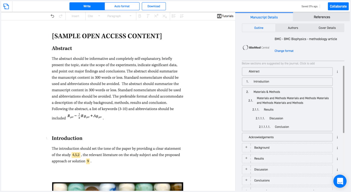
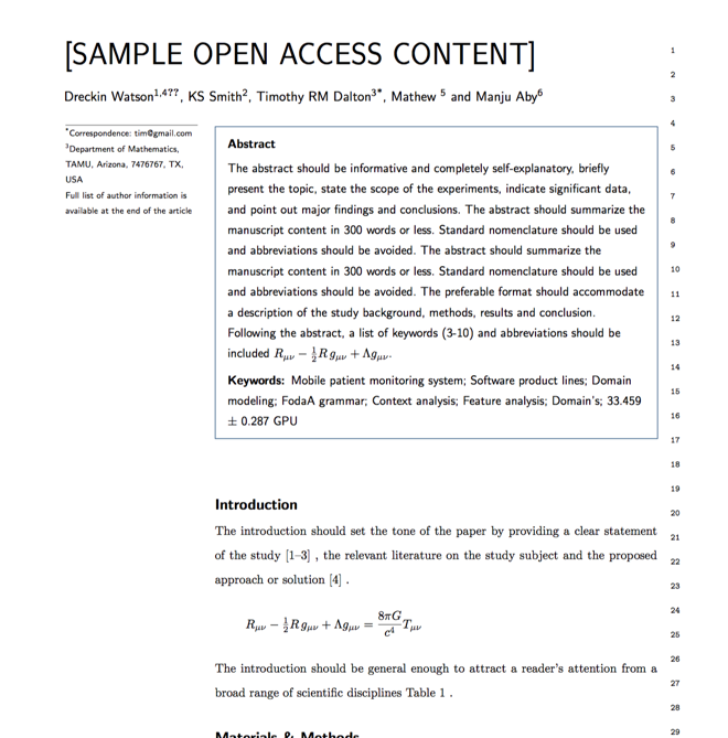
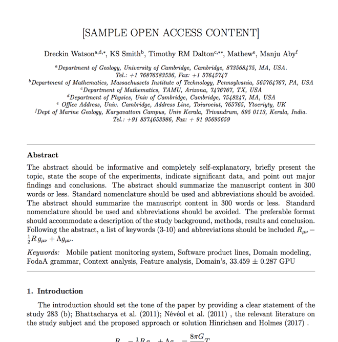
With SciSpace, you do not need a word template for Microscopy and Microanalysis.

It automatically formats your research paper to Cambridge University Press formatting guidelines and citation style.

You can download a submission ready research paper in pdf, LaTeX and docx formats.

Time comparison

Time taken to format a paper and Compliance with guidelines

Plagiarism Reports via Turnitin

SciSpace has partnered with Turnitin, the leading provider of Plagiarism Check software.

Using this service, researchers can compare submissions against more than 170 million scholarly articles, a database of 70+ billion current and archived web pages. How Turnitin Integration works?

Turnitin Stats
Publisher Logos

Freedom from formatting guidelines

One editor, 100K journal formats – world's largest collection of journal templates

With such a huge verified library, what you need is already there.

publisher-logos

Easy support from all your favorite tools

Microscopy and Microanalysis format uses unsrt citation style.

Automatically format and order your citations and bibliography in a click.

SciSpace allows imports from all reference managers like Mendeley, Zotero, Endnote, Google Scholar etc.

Frequently asked questions

1. Can I write Microscopy and Microanalysis in LaTeX?

Absolutely not! Our tool has been designed to help you focus on writing. You can write your entire paper as per the Microscopy and Microanalysis guidelines and auto format it.

2. Do you follow the Microscopy and Microanalysis guidelines?

Yes, the template is compliant with the Microscopy and Microanalysis guidelines. Our experts at SciSpace ensure that. If there are any changes to the journal's guidelines, we'll change our algorithm accordingly.

3. Can I cite my article in multiple styles in Microscopy and Microanalysis?

Of course! We support all the top citation styles, such as APA style, MLA style, Vancouver style, Harvard style, and Chicago style. For example, when you write your paper and hit autoformat, our system will automatically update your article as per the Microscopy and Microanalysis citation style.

4. Can I use the Microscopy and Microanalysis templates for free?

Sign up for our free trial, and you'll be able to use all our features for seven days. You'll see how helpful they are and how inexpensive they are compared to other options, Especially for Microscopy and Microanalysis.

5. Can I use a manuscript in Microscopy and Microanalysis that I have written in MS Word?

Yes. You can choose the right template, copy-paste the contents from the word document, and click on auto-format. Once you're done, you'll have a publish-ready paper Microscopy and Microanalysis that you can download at the end.

6. How long does it usually take you to format my papers in Microscopy and Microanalysis?

It only takes a matter of seconds to edit your manuscript. Besides that, our intuitive editor saves you from writing and formatting it in Microscopy and Microanalysis.

7. Where can I find the template for the Microscopy and Microanalysis?

It is possible to find the Word template for any journal on Google. However, why use a template when you can write your entire manuscript on SciSpace , auto format it as per Microscopy and Microanalysis's guidelines and download the same in Word, PDF and LaTeX formats? Give us a try!.

8. Can I reformat my paper to fit the Microscopy and Microanalysis's guidelines?

Of course! You can do this using our intuitive editor. It's very easy. If you need help, our support team is always ready to assist you.

9. Microscopy and Microanalysis an online tool or is there a desktop version?

SciSpace's Microscopy and Microanalysis is currently available as an online tool. We're developing a desktop version, too. You can request (or upvote) any features that you think would be helpful for you and other researchers in the "feature request" section of your account once you've signed up with us.

10. I cannot find my template in your gallery. Can you create it for me like Microscopy and Microanalysis?

Sure. You can request any template and we'll have it setup within a few days. You can find the request box in Journal Gallery on the right side bar under the heading, "Couldn't find the format you were looking for like Microscopy and Microanalysis?”

11. What is the output that I would get after using Microscopy and Microanalysis?

After writing your paper autoformatting in Microscopy and Microanalysis, you can download it in multiple formats, viz., PDF, Docx, and LaTeX.

12. Is Microscopy and Microanalysis's impact factor high enough that I should try publishing my article there?

To be honest, the answer is no. The impact factor is one of the many elements that determine the quality of a journal. Few of these factors include review board, rejection rates, frequency of inclusion in indexes, and Eigenfactor. You need to assess all these factors before you make your final call.

13. What is Sherpa RoMEO Archiving Policy for Microscopy and Microanalysis?

SHERPA/RoMEO Database

We extracted this data from Sherpa Romeo to help researchers understand the access level of this journal in accordance with the Sherpa Romeo Archiving Policy for Microscopy and Microanalysis. The table below indicates the level of access a journal has as per Sherpa Romeo's archiving policy.

RoMEO Colour Archiving policy
Green Can archive pre-print and post-print or publisher's version/PDF
Blue Can archive post-print (ie final draft post-refereeing) or publisher's version/PDF
Yellow Can archive pre-print (ie pre-refereeing)
White Archiving not formally supported
FYI:
  1. Pre-prints as being the version of the paper before peer review and
  2. Post-prints as being the version of the paper after peer-review, with revisions having been made.

14. What are the most common citation types In Microscopy and Microanalysis?

The 5 most common citation types in order of usage for Microscopy and Microanalysis are:.

S. No. Citation Style Type
1. Author Year
2. Numbered
3. Numbered (Superscripted)
4. Author Year (Cited Pages)
5. Footnote

15. How do I submit my article to the Microscopy and Microanalysis?

It is possible to find the Word template for any journal on Google. However, why use a template when you can write your entire manuscript on SciSpace , auto format it as per Microscopy and Microanalysis's guidelines and download the same in Word, PDF and LaTeX formats? Give us a try!.

16. Can I download Microscopy and Microanalysis in Endnote format?

Yes, SciSpace provides this functionality. After signing up, you would need to import your existing references from Word or Bib file to SciSpace. Then SciSpace would allow you to download your references in Microscopy and Microanalysis Endnote style according to Elsevier guidelines.

Fast and reliable,
built for complaince.

Instant formatting to 100% publisher guidelines on - SciSpace.

Available only on desktops 🖥

No word template required

Typset automatically formats your research paper to Microscopy and Microanalysis formatting guidelines and citation style.

Verifed journal formats

One editor, 100K journal formats.
With the largest collection of verified journal formats, what you need is already there.

Trusted by academicians

I spent hours with MS word for reformatting. It was frustrating - plain and simple. With SciSpace, I can draft my manuscripts and once it is finished I can just submit. In case, I have to submit to another journal it is really just a button click instead of an afternoon of reformatting.

Andreas Frutiger
Researcher & Ex MS Word user
Use this template